All vertebrates have the kidneys, a pair of bean-shaped organs. They maintain normal electrolyte levels, control blood pressure, and eliminate waste from the body. Kidney is one of the body’s most crucial organs. History records that the ancient Egyptians only preserved the brain and kidneys before embalming a body, implying that they were of greater worth than other organs.
Location
The kidneys are situated on either side of the spine, directly below the rib cage. To pave way for the liver, the right kidney is often slightly lower than the left kidney. The kidneys measure around 3 centimeters (cm) in thickness, 6 cm in width, and 12 cm in length. The typical weight of a male’s right and left kidneys is about 129 grams (g) and 137 g, respectively. These organs typically weigh 108 g for the right kidney and 116 g for the left kidney in females.
Structure
The kidneys are two organs that resemble beans and measure about the size of a fist. Each kidney is encased in a hard, fibrous renal capsule that supports the delicate tissue inside. Two additional layers of fat provide further defense. The kidneys are located on top of the adrenal glands.
There are several pyramid-shaped lobes inside the kidneys. Each kidney has an inner renal medulla and an outer renal cortex. These portions are connected by nephrons. A filter called the glomerulus and a tubule are both parts of a nephron.
Blood is filtered in the glomerulus after it passes through the kidneys’ renal arteries and veins. Despite being quite small, the kidneys get 20–25% of the heart’s output. The tubule eliminates waste, which is then converted to urine, and returns vital components to the circulation. Through the ureter, a tube that connects to the bladder, the kidneys expel urine.
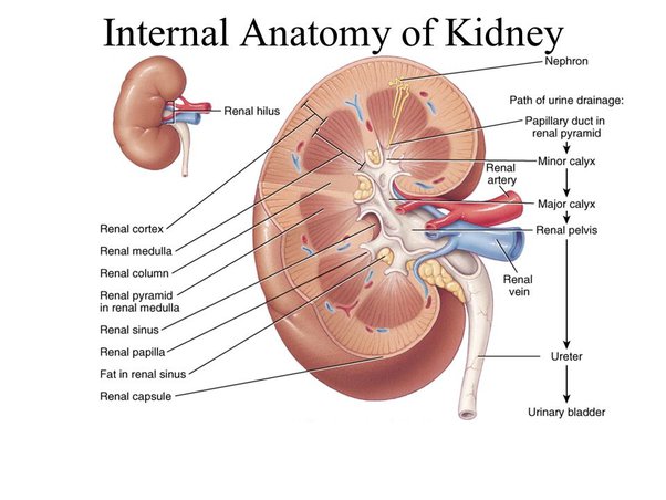
Parts of the Kidney
1. Nephrons
The most central part of each kidney is the nephron. They ingest blood, digest nutrients, and assist in excreting waste from filtered blood. There are roughly 1 million nephrons per kidney. Each has a unique collection of internal structures.
2. Renal Corpuscle
Blood flows into the renal corpuscle, also known as a Malpighian body, after entering a nephron. Two more structures can be seen in the renal corpuscle:
- The glomerulus: This group of capillaries is responsible for absorbing protein from blood as it passes through the renal corpuscle.
- Bowman’s capsule: Capsular urine, the leftover urine, enters the renal tubules through the Bowman capsule.
3. Renal Tubules
The Bowman capsule is followed by a series of tubes called the renal tubules, which terminate at collecting ducts.
There are various sections to each tubule:
- Proximal convoluted tubule: Reabsorbing glucose, salt, and water into the circulation is what this area does.
- Loop of Henle: Potassium, chloride, and sodium are further absorbed into the circulation in this region.
- Distal convoluted tubule: More sodium is absorbed into the circulation in this area, along with potassium and acid. The fluid is diluted and filled with urea by the time it reaches the tubule’s end. Protein metabolism produces urea, which is excreted in the urine.
4. Renal Cortex
The outside of the kidney is known as the renal cortex. It has convoluted tubules and the glomerulus. The renal capsule, a layer of fatty tissue, encircles the renal cortex on both sides. The renal cortex and capsule together house and safeguard the inner structures of the
5. Renal Medulla
The kidney’s smooth inner tissue is known as the renal medulla. It contains the loop of Henle as well as renal pyramids.
6. Renal Pyramids
Strings of nephrons and tubules are seen inside the tiny structures known as renal pyramids. The kidney receives fluid through these tubules. The fluid then flows away from the nephrons and in the direction of the inner kidney structures that store and transport urine.
7. Collecting Ducts
The renal medulla has a collecting duct at the end of each nephron. Filtered fluids leave the nephrons at this point. The fluid continues on to its last destinations in the renal pelvis after entering the collecting duct.
8. Renal Pelvis
In the deepest region of the kidney, there is a funnel-shaped cavity known as the renal pelvis. It serves as a pathway for fluid on its way to the bladder
9. Calyces
The calyces are located in the first segment of the renal pelvis. Before fluid enters the bladder, it is collected in these tiny cup-shaped pockets. Urine is also created at this location from surplus fluid and waste.
10. Hilum
The kidney’s inner edge has a small aperture called the hilum, which is where the kidney curls inward to give it its distinctive bean-like form. It also accommodates the renal pelvis and the:
- Renal artery. This brings oxygenated blood from the heart to the kidney for filtration.
- Renal vein. This carries filtered blood from the kidneys back to the heart.
11. Ureter
A muscular tube called the ureter forces urine into the bladder, where it collects and is excreted from the body.
Functions of the Kidney
Homeostasis
The maintenance of homeostasis is the kidneys’ primary function. They control fluid levels, electrolyte balance, and other elements that ensure the body’s stable and comfortable interior environment.
Excretion of Waste
Numerous waste items are removed by the kidneys and eliminated in the urine. Among the main substances the kidneys filter are:
- Urea, which results from the breakdown of proteins.
- Uric acid from the breakdown of nucleic acids.
- Drugs and their metabolites.
Reabsorption of nutrients
The kidneys recycle fluids tubules draw nutrients from the blood and deliver them to areas of the body where they will support health the most. To sustain homeostasis, they also reabsorb other substances. Reabsorbed products consist of:
- Glucose
- Amino acids
- Bicarbonate
- Water
- Phosphate
- Chloride, sodium, magnesium, and potassium ions
Maintaining pH
The pH range that is suitable for humans is 7.35 to 7.45. The body assumes a state of acidemia or alkalemia, respectively, at levels below or above this range. Proteins and enzymes degrade in these conditions and stop working. In dire circumstances, this may be lethal.
The lungs and kidneys aid in maintaining the body’s pH balance. The lungs accomplish this by regulating the blood’s carbon dioxide levels. By generating and reabsorbing bicarbonate from urine, which helps neutralize acids, the kidneys regulate pH.
If the pH is bearable, the kidneys can retain bicarbonate and can release it if the acidity level increases. They can excrete acid, which they can then use to make new bicarbonate.
Osmolality Control
Osmolality, or the ratio of bodily fluids to minerals, is a measurement of the electrolyte-water balance in the body. The main factor causing electrolyte imbalance is dehydration.
When blood plasma osmolality increases, the brain’s hypothalamus reacts by sending a signal to the pituitary gland. Antidiuretic hormone is released by this gland (ADH). The kidney changes in response to ADH in a number of ways, including:
- Increasing urine concentration.
- Increasing water reabsorption.
- Reopening portions of the collecting duct that water cannot normally enter, allowing water back into the body.
- Retaining urea in the medulla of the kidney rather than excreting it, as this compound draws in water.
Regulation of Blood Pressure
When blood pressure needs to be adjusted, the kidneys are in charge of slower changes.
By altering the fluid around cells, they influence the long-term pressure in the arteries. The medical term for this fluid is extracellular fluid. Following the production of a vasoconstrictor called angiotensin II, these fluid shifts take place. Hormones called vasoconstrictors make blood arteries constrict.
These hormones contribute to an increase in salt, or sodium chloride, absorption by the kidneys. Blood pressure rises as a result of this absorption, which also expands the extracellular fluid compartment. Chronic kidney impairment can result from anything that affects blood pressure, such as heavy alcohol use, smoking, and obesity.
Secretion of Energetic Substances
Several significant substances are released by the kidneys, including:
- Erythropoiesis: Red blood cell production, or erythropoiesis, is regulated by the hormone erythropoietin. Erythropoietin is also produced by the liver, but in adulthood, the kidneys are the primary source.
- Renin: This enzyme aids in controlling artery growth as well as blood plasma, lymph, and interstitial fluid levels. White blood cells, which promote immune function, are found in the fluid known as lymph, and interstitial fluid makes up the majority of extracellular fluid.
- Calcitriol: The hormone-active metabolite of vitamin D is called calcitriol. It increases the capacity of the intestines to absorb calcium as well as the kidneys’ capacity to reabsorb phosphate.